|
Visual Snow Syndrome Improves With Modulation of Resting-State Functional MRI Connectivity After Mindfulness-Based Cognitive Therapy: An Open-Label Feasibility Study
Wong, Sui H. MD, FRCP; Pontillo, Giuseppe PhD; Kanber, Baris PhD; Prados, Ferran PhD; Wingrove, Janet PhD, CPsychol; Yiannakas, Marios PhD; Davagnanam, Indran FRCR; Gandini Wheeler-Kingshott, Claudia A. M. PhD; Toosy, Ahmed T. PhD, FRCP Background: Visual snow syndrome (VSS) is associated with functional connectivity (FC) dysregulation of visual networks (VNs). We hypothesized that mindfulness-based cognitive therapy, customized for visual symptoms (MBCT-vision), can treat VSS and modulate dysfunctional VNs. Methods: An open-label feasibility study for an 8-week MBCT-vision treatment program was conducted. Primary (symptom severity; impact on daily life) and secondary (WHO-5; CORE-10) outcomes at Week 9 and Week 20 were compared with baseline. Secondary MRI outcomes in a subcohort compared resting-state functional and diffusion MRI between baseline and Week 20. Results: Twenty-one participants (14 male participants, median 30 years, range 22–56 years) recruited from January 2020 to October 2021. Two (9.5%) dropped out. Self-rated symptom severity (0–10) improved: baseline (median [interquartile range (IQR)] 7 [6–8]) vs Week 9 (5.5 [3–7], P = 0.015) and Week 20 (4 [3–6], P < 0.001), respectively. Self-rated impact of symptoms on daily life (0–10) improved: baseline (6 [5–8]) vs Week 9 (4 [2–5], P = 0.003) and Week 20 (2 [1–3], P < 0.001), respectively. WHO-5 Wellbeing (0–100) improved: baseline (median [IQR] 52 [36–56]) vs Week 9 (median 64 [47–80], P = 0.001) and Week 20 (68 [48–76], P < 0.001), respectively. CORE-10 Distress (0–40) improved: baseline (15 [12–20]) vs Week 9 (12.5 [11–16.5], P = 0.003) and Week 20 (11 [10–14], P = 0.003), respectively. Within-subject fMRI analysis found reductions between baseline and Week 20, within VN-related FC in the i) left lateral occipital cortex (size = 82 mL, familywise error [FWE]-corrected P value = 0.006) and ii) left cerebellar lobules VIIb/VIII (size = 65 mL, FWE-corrected P value = 0.02), and increases within VN-related FC in the precuneus/posterior cingulate cortex (size = 69 mL, cluster-level FWE-corrected P value = 0.02). Conclusions: MBCT-vision was a feasible treatment for VSS, improved symptoms and modulated FC of VNs. This study also showed proof-of-concept for intensive mindfulness interventions in the treatment of neurological conditions. Published online 2023 May 30. doi: 10.2217/cnc-2023-0006
PMCID: PMC10498816 PMID: 37711469 Neuro-optometric treatment for visual snow syndrome: recent advances Kenneth J Ciuffreda, Barry Tannen, Daniella Rutner, and MH Esther Han "In conclusion, the condition of visual snow syndrome, which is found is found in approximately 2% of the general population, has received considerable attention over the past decade by both clinicians and researchers. Fortunately, two neuro-optometric rehabilitative approaches have benefitted most patients. This has included colored filters and eye movement procedures to reduce the perception of the visual snow and some of the other related visual disturbances, such as light sensitivity." In this informative blog article, Visual Snow Syndrome Glasses for Symptom Relief, Jen Ambrose, founder of the Eye on Vision Foundation, shares her personal journey with Visual Snow Syndrome (VSS) and offers valuable advice to others living with the condition. Exploring the challenges of VSS and its impact on daily life, she delves into the power of Visual Snow Syndrome glasses, specifically FL-41 glasses, in managing light sensitivity and reducing visual disturbances. With a message of hope and solidarity, Jen inspires others to embrace their unique journey with VSS and find comfort in knowing they are not alone.
In this heartfelt and personal blog article, Living with Visual Snow Syndrome, I share my journey of living with Visual Snow Syndrome (VSS). I delve into the challenges I faced, including misdiagnosis and the impact on daily life. Through my experiences, I highlight the importance of finding support in online communities and developing coping mechanisms. I emphasize the significance of self-care, building a support network, and seeking empowerment. By sharing my story, I hope to inspire others living with VSS to find hope, strength, and resilience in their own journeys.
The efficacy of neuro-optometric visual rehabilitation therapy in patients with visual snow syndrome1/24/2023
Terry Tsang, Charles Shidlofsky, Vanessa Mora "Conclusion: Our results suggest that patients with VSS experience improvement in QOL in as little as 6 weeks, with further improvement by 12 weeks of NORT. This suggests NORT is an effective treatment option for managing the condition and improving QOL in patients with VSS, although a reduction in specific symptoms has yet to be demonstrated. This study provides justification that NORT warrants further investigation on VSS symptom reduction."
Full article can be found by clicking here Barry Tannen, OD, FCOVD, FAAO Jacob Brown, OD Kenneth J. Ciuffreda, OD, PhD, FCOVD, FAAO, FARVO Noah M. Tannen, OD, FCOVD, FAAO A small excerpt from the article with full PDF attached This is the first paper to demonstrate successful neuro-optometric rehabilitation (NOR) in a clinical population diagnosed with VSS and its wide array of unique visual symptoms. It included the use of: (1) chromatic tints/filters to reduce the perceived intensity and frequency of the VS and many of the other abnormal visual phenomena (e.g., palinopsia)reported in those with VSS; these filters reduce the overall illumination of the visual field, more so in the specific “offensive” spectral band, which typically appears to be in the blue region of the visible spectrum.14 (2) Saccadic tracking to reduce the perceived intensity and frequency of the palinopsia, which has been speculated to occur due to a disinhibition/ hypersensitivity phenomenon related to saccadic suppression;6,7 the training appears to reestablish/reset a more normal level of saccadic suppression, such that its smeared, afterimage perception during a saccade is inhibited once again. In addition, and somewhat serendipitously, this is the first study to detect, diagnose, and treat the multitude of versional oculomotor deficits (i.e., OMD) that appear to be present with a very high frequency of occurrence in our sample (~60%), and likely present more generally.15,16 This is fruitful territory for future investigations (see later Discussion). We have found that common optometric diagnostic tests (DEM, Visagraph, and direct observation of eye movements) combine to allow for the diagnosis of oculomotor dysfunction.
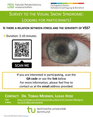 VSS and Stress
Lukas Hose reached out to Eye on Vision Foundation seeking assistance in reaching more members of the Visual Snow community. Please read the following message for more information. Dear VSS-Community. As long as I can remember, I have a colorful veil of flickering dots in my visual field. As I shut my eyes, the flickering itself is accompanied by several other visual symptoms - like a negative image of light sources deforming in front of my inner eye, pulsating and changing colours. At the age of 16 I approached other people about it and - surprise - not everyone perceives the world with a snow-like flickering. This ignited my search for an explanation. Years passed, and as soon as I had the idea to compare the flickering with the snow of an analog TV, I found the term “visual snow”. With a name for my visual phenomena, I was able to connect with other people affected, and learned that VS is different for everyone. However, a common advice for new members in social media groups said to "avoid stress" and to not concentrate on the symptoms, as this worsens the experience. This rang a bell. For many, the onset of VSS is a very traumatic experience, making them feel anxious and powerless. I could empathize with them but in my own experience, VSS is nothing to be afraid of and that it is possible to ease the symptoms if you grow with them. Today, I created a small survey about a possible relation between stress and VSS. Maybe my results can change the outcome for some people. This would mean the world to me. So if you want to help me with my thesis, please participate in my survey. Thank you very much and all the best! Lukas https://umfragen.tu-dortmund.de/index.php/921391?newtest=Y&lang=en Visual snow syndrome is probably not mediated by CGRP: A case series
Stefan Evers, Dagny Holle-Lee, Christoph J Schankin First Published May 25, 2022 https://doi.org/10.1177/03331024221099220 Visual snow syndrome is a phenomenon for which no effective treatment is known. It is highly comorbid with migraine, therefore we performed a retrospective chart review of patients with visual snow syndrome treated with a monoclonal antibody against calcitonin gene related peptide or its receptor. We enrolled 15 patients with visual snow syndrome who received at least once a monoclonal antibody against calcitonin gene related peptide or its receptor. None of the patients reported relief of visual snow syndrome whereas those patients with comorbid migraine reported a very good efficacy of the antibody against the migraine headache but not against the migraine aura. The data suggest that visual snow syndrome is not mediated by calcitonin gene related peptide in a relevant way and that the calcitonin gene related peptide receptor is not involved in the network underlying the visual snow syndrome. Myrte Strik, Meaghan Clough, Emma J Solly, Rebecca Glarin, Owen B White, Scott C Kolbe, Joanne Fielding Brain Communications, Volume 4, Issue 4, 2022, fcac164, https://doi.org/10.1093/braincomms/fcac164 Conclusion It is clear that visual snow syndrome is a disorder of the central nervous system. However, the underlying pathophysiological mechanisms remains elusive. Here, we reveal no evidence of gross morphometry changes in the visual snow syndrome brain, but widespread changes in the microstructure of the GM, the most notable of these occurring in caudal regions including the occipital cortex. None of these changes are directly associated with the co-occurrence of migraine. While we were unable to determine the specific brain tissue that underlies microstructural changes, they do focus further investigations, contributing significantly to our understanding of visual snow syndrome. * GM refers to gray matter Download and read the full PDF version of the study
The Psychiatric Symptomatology of Visual Snow Syndrome
Emma J. Solly Meaghan Clough Paige Foletta Owen B. White Joanne Fielding Conclusion: Psychiatric symptoms are highly prevalent in patients with VSS and are associated with increased visual symptom severity and reduced quality of life. Importantly, patients with lifelong VSS reported lower levels of distress and milder self-ratings of visual symptoms compared to patients with a later onset, while being equally likely to experience psychiatric symptoms. This suggests that the psychiatric symptoms of VSS are not solely due to distress caused by visual symptoms. While no consistently effective treatments are available for the visual symptomology of VSS, psychiatric symptoms offer an avenue of treatment that is likely to significantly improve patient quality of life and ability to cope with visual symptoms. Full article: https://www.frontiersin.org/articles/10.3389/fneur.2021.703006/full |
UpdateRecent update on research. Archives
November 2023
Categories |
||||
 RSS Feed
|
|
|