|
We are pleased to announce that the Eye on Vision Foundation was able to donate $30,000 towards Dr. Schankin's latest VSS research project.
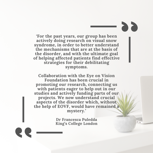
Dr. Schankin leads the University Headache Clinic at Inselspital, within its Neurology Department. His academic journey began at the University of Heidelberg and extended to Columbia University in New York. Further enhancing his expertise, he pursued research at the University of York's Biology Department in the UK. A significant milestone in his career was his collaboration with Dr. Peter Goadsby at the University of California, San Francisco's Headache Center. This experience not only broadened Dr. Schankin's expertise, particularly in Visual Snow Syndrome, various headache types including rare and secondary headaches, and the intricacies of migraine and related visual disturbances, but was also the beginning of the collaboration between Dr. Schankin, Prof. Goadsby and the Eye on Vision Foundation. Dr. Schankin is an esteemed member of several national and international neurological societies. His exceptional contributions to the field have been recognized with numerous awards, notably the Harold G. Wolff Lecture Award from the American Headache Society and the Wolffram Prize from the German Migraine and Headache Society. In a new venture, Dr. Schankin is collaborating with Dr. Antonia Klein, a talented researcher and Neurology Resident at Inselspital, Bern University Hospital. They are embarking on a pioneering study focusing on Visual Snow Syndrome, promising to add valuable insights to the field. Researchers from King’s College London have used a novel approach to show that the patterns of activity in two brain chemical systems - glutamate and serotonin – are different in people with visual snow syndrome compared to those without the condition.
Visual snow syndrome is characterised by a continuous visual disturbance in which people see static, flickering dots, and flashing lights – this happens when their eyes are both open and closed. It affects about 2 -3% of the world’s population and it can be debilitating, impacting vision, hearing, thinking, sensory processing, and quality of life. The study was part-funded by the National Institute for Health and Care Research (NIHR) Maudsley Biomedical Research Centre, and supported by the Visual Snow Initiative and the Eye on Vision Foundation. Researchers used existing scanning data from 24 patients with visual snow syndrome (VSS), 25 migraine patients and 24 healthy people with no VSS or migraine history. The brain scans were conducted at the NIHR King’s Clinical Research Facility. Tackling the lack of understanding around the biology of the condition Currently there is no medication that is effective at helping people with VSS and the search for treatment has been hindered by lack of knowledge around the biology of the condition. Previous scanning studies in people with VSS have shown alterations in different regions of the brain and how these interact, however, there has been no research looking into the possible molecular changes that could underlie the condition. Further research into this area could provide insight into potential biological targets for treatment. First author on the study and Senior Clinical Research Fellow at the Institute of Psychiatry, Psychology & Neuroscience (IoPPN), King’s College London, Dr Francesca Puledda said: “Previous research has given us some insight into the brain regions involved in visual snow syndrome but our study is the first to identify the brain chemistry that could underlie the condition and other sensory experiences such as migraine aura. Until now there have been no effective treatments for VSS and importantly our research has confirmed that VSS is a neurological disorder with a chemical basis that could be used as a target for development of treatments.” Mapping brain activity using innovative scanning techniques Using a new approach that was developed by researchers at the NIHR Maudsley BRC, the study combines information from positron emission tomography (PET) imaging information on the distribution of different chemical receptors in the brain with functional Magnetic Resonance Imaging (fMRI) analysis of the interaction of activity between different regions of the brain. Using this Receptor-Enriched Analysis of Functional Connectivity by Targets (REACT) approach researchers can extract a map of activity of brain chemicals across the different brain areas. The five brain chemicals examined in this study were noradrenaline, dopamine, serotonin, glutamate and GABA. Differences found in the activity of glutamate and serotonin networks Researchers found that in patients with VSS there were particular differences in the activity of glutamate and serotonin networks in specific areas of the brain. There was less synchronised activity (or functional connectivity) in the glutamate networks in the anterior cingulate cortex (ACC) in those with VCC compared to healthy controls and those with migraine. The ACC is a hub for thinking and top-down control over sensory inputs and the different pattern of activity could represent an interruption in the filtering and integration of visual information. Analysis also showed that VSS patients had reduced functional connectivity in the serotonin networks of the visual cortex, insula, temporal pole and orbitofrontal areas of the brains compared to healthy controls. This reduced connectivity in serotonin networks was also seen in migraine patients with aura suggesting a biological link between VSS and aura. The findings suggest that serotonin activity in VSS patients may be influencing the integration of complex sensory information. The results did not find any differences for the other brain chemicals that were investigated in the study. Colorado rTMS study enrollment has concluded in CO, and data analysis will being in January 20239/18/2022
 Colorado rTMS study enrollment has concluded in CO, and data analysis will being in January 2023.
Study details: The study will gather information about the treatment of up to 10 people with VS using rTMS. Treated participants will undergo 10 sessions of rTMS administered 5 times a week over 2 weeks. All visits will take place in the University of Colorado School of Medicine NeuroMag/Transcranial Magnetic Stimulation laboratory on University of Colorado Anschutz Medical Campus. The specific aims for this feasibility study include: Determine whether any participant experiences untoward effects of rTMS in the setting of visual snow syndrome and determine the potential drop-out rate of larger study. Determine the performance of a novel scale (Colorado Visual Snow Scale) and two three psychophysical visual processing tasks Determine the standard deviation and test-retest reliability for the novel scale and two visual processing tasks Determine whether the visual processing tasks perform similar to performance found by the developer Describe changes in outcome measures between pre- and post-treatment with rTMS Study overseen by Dr. Pelak.  With this project, we aim at reducing VSS symptoms (i.e. visual snow, palinopsia, entoptic
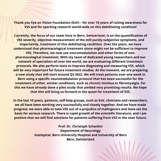
phenomena, photophobia, nyctalopia) using transcranial electric stimulation (tACS). Targeted treatment approach doing neuromodulation planned at Inselspital Bern Start will be in autumn 2022 We have promising preliminary data from a previous study performed with support by EOV foundation Treatment will be over one week a in Bern Lead researchers are Prof. Dr. med. Christoph Schankin and Antonia Klein, MD Study has partial funding but would need 90000 USD for completion of the study Donations are accepted at EyeOnVision.org Antonia Klein MD, Christoph J. Schankin MD First published: 27 September 2021 https://doi.org/10.1111/head.14213 Abstract Objective The aim of this narrative review is to explore the relationship between visual snow syndrome (VSS), migraine, and a group of other perceptual disorders. Background VSS is characterized by visual snow and additional visual and nonvisual disturbances. The clinical picture suggests a hypersensitivity to internal and external stimuli. Imaging and electrophysiological findings indicate a hyperexcitability of the primary and secondary visual areas of the brain possibly due to an impairment of inhibitory feedback mechanisms. Migraine is the most frequent comorbidity. Epidemiological and clinical studies indicate that other perceptual disorders, such as tinnitus, fibromyalgia, and dizziness, are associated with VSS. Clinical overlaps and parallels in pathophysiology might exist in relation to migraine. Methods We performed a PubMed and Google Scholar search with the following terms: visual snow syndrome, entoptic phenomenon, fibromyalgia, tinnitus, migraine, dizziness, persistent postural-perceptual dizziness (PPPD), comorbidities, symptoms, pathophysiology, thalamus, thalamocortical dysrhythmia, and salience network. Results VSS, fibromyalgia, tinnitus, and PPPD share evidence of a central disturbance in the processing of different stimuli (visual, somatosensory/pain, acoustic, and vestibular) that might lead to hypersensitivity. Imaging and electrophysiological findings hint toward network disorders involving the sensory networks and other large-scale networks involved in the management of attention and emotional processing. There are clinical and epidemiological overlaps between these disorders. Similarly, migraine exhibits a multisensory hypersensitivity even in the interictal state with fluctuation during the migraine cycle. All the described perceptual disorders are associated with migraine suggesting that having migraine, that is, a disorder of sensory processing, is a common link. Conclusion VSS, PPPD, fibromyalgia, and chronic tinnitus might lie on a spectrum of perceptual disorders with similar pathophysiological mechanisms and the common risk factor migraine. Understanding the underlying network disturbances might give insights into how to improve these currently very difficult to treat conditions. To view full article click here
Francesca Puledda, Muriel Bruchhage, Owen O'Daly, Dominic Ffytche, Steven C.R. Williams, Peter J. Goadsby First published August 5, 2020, DOI: https://doi.org/10.1212/WNL.0000000000010530 Abstract Objective To determine whether regional gray and white matter differences characterize the brain of patients with visual snow syndrome, a newly defined neurologic condition, we used a voxel-based morphometry approach. Methods In order to investigate whole brain morphology directly, we performed an MRI study on patients with visual snow syndrome (n = 24) and on age- and sex-matched healthy volunteers (n = 24). Voxel-based morphometry was used to determine volumetric differences in patients with visual snow. We further analyzed cerebellar anatomy directly using the high-resolution spatially unbiased atlas template of the cerebellum. Results Compared to healthy controls, patients with visual snow syndrome had increased gray matter volume in the left primary and secondary visual cortices, the left visual motion area V5, and the left cerebellar crus I/lobule VI area. These anatomical alterations could not be explained by clinical features of the condition. Conclusion Patients with visual snow syndrome have subtle, significant neuroanatomical differences in key visual and lateral cerebellar areas, which may in part explain the pathophysiologic basis of the disorder. To access the full article please download the PDF below or download here
Disrupted connectivity within visual, attentional and salience networks in the visual snow syndrome1/4/2022
Francesca Puledda, Owen O'Daly, Christoph Schankin, Dominic Ffytche, Steven CR Williams, Peter J Goadsby First published: 15 January 2021 https://doi.org/10.1002/hbm.25343 Abstract Here we investigate brain functional connectivity in patients with visual snow syndrome (VSS). Our main objective was to understand more about the underlying pathophysiology of this neurological syndrome. Twenty-four patients with VSS and an equal number of gender and age-matched healthy volunteers attended MRI sessions in which whole-brain maps of functional connectivity were acquired under two conditions: at rest while watching a blank screen and during a visual paradigm consisting of a visual-snow like stimulus. Eight unilateral seed regions were selected a priori based on previous observations and hypotheses; four seeds were placed in key anatomical areas of the visual pathways and the remaining were derived from a pre-existing functional analysis. The between-group analysis showed that patients with VSS had hyper and hypoconnectivity between key visual areas and the rest of the brain, both in the resting state and during a visual stimulation, compared with controls. We found altered connectivity internally within the visual network; between the thalamus/basal ganglia and the lingual gyrus; between the visual motion network and both the default mode and attentional networks. Further, patients with VSS presented decreased connectivity during external sensory input within the salience network, and between V5 and precuneus. Our results suggest that VSS is characterised by a widespread disturbance in the functional connectivity of several brain systems. This dysfunction involves the pre-cortical and cortical visual pathways, the visual motion network, the attentional networks and finally the salience network; further, it represents evidence of ongoing alterations both at rest and during visual stimulus processing. Please click here to read full article
|
Eye On Vision FoundationCollaborating with and funding researchers since 2006. Archives
November 2023
Categories |
||||
 RSS Feed
|
|